Macromolecules play key roles in the structure and function of cells. Understanding macromolecules helps us learn how organisms grow, develop, and maintain their systems. Beyond the body, they are used in medical treatments, nutrition and biotechnology. Use this resource to learn about the types of macromolecules.
Macromolecules are large, complex molecules essential for life. They include carbohydrates, proteins, lipids, and nucleic acids. Each type has a different role within a cell. For example, proteins help build and repair tissues, while nucleic acids like DNA store genetic information.
Carbohydrates
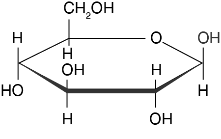
Carbohydrates are macromolecules made up of simple sugar monomers (units) called monosaccharides (“mono” meaning “one” and “saccharide” meaning “sugar”). These can be linked together to form repeating polymers called disaccharides (“di” meaning “two”, i.e. two sugar monomers) or polysaccharides (“poly” meaning “many”, i.e. many sugar monomers). Glucose
There are three different types of carbohydrates:
sugars – such as glucose and fructose in fruits, vegetables and honey
starches – such as amylose and amylopectin found in potatoes, rice and corn
fibre – such as cellulose, oligosaccharides and chitins found in leafy vegetables, grains and crustaceans.
Carbohydrates are composed of carbon (\(\ce{C}\)) atoms, hydrogen (\(\ce{H}\)) atoms and oxygen (\(\ce{O}\)) atoms. The “carbo” part of its name refers to the carbon atoms and the “hydrate” part refers to the presence of water (\(\ce{H2}\)). For example, glucose has the formula \(\ce{C6H12O6}\) and amylose \(\ce{(C6H10O5)_{n}}\), where \(n\) represents the number of repeating glucose units.
In living organisms, carbohydrates are the primary source of energy. They are crucial for cell function and metabolism.
Formation of carbohydrates
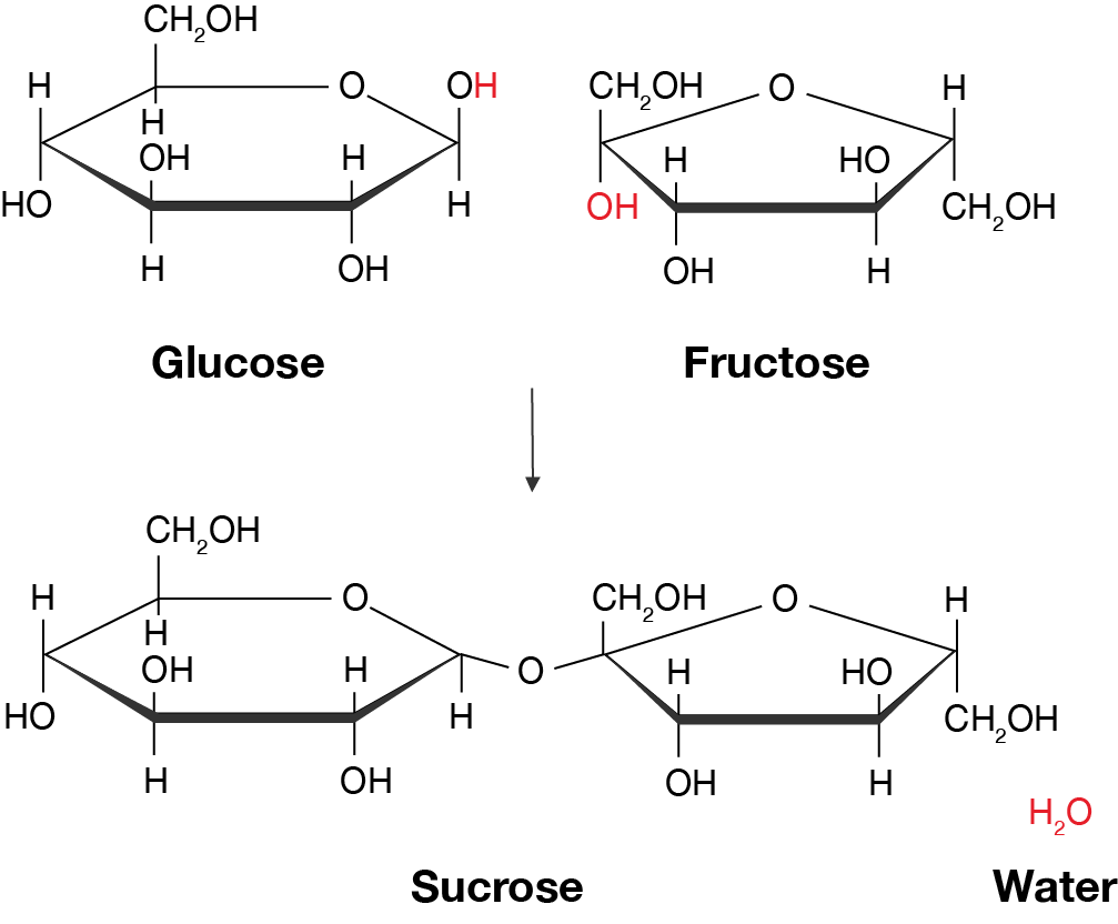
Carbohydrates are formed through a condensation reaction between monosaccharides. This involves the \(\ce{OH}\) groups from two monomers coming together and reacting to form a glycosidic bond. A water molecule \(\ce{H2O}\) is released from the reaction; this is a key characteristic of condensation reactions.
An example of this is the reaction between glucose and fructose monomers to form sucrose and a water molecule. Sucrose is a disaccharide because it consists of two monosaccharides. Condensation reaction between gluose and fructose to form sucrose and a water molecule
Carbohydrate formation occurs primarily in plants through photosynthesis. Animals then obtain carbohydrates from their diets, i.e. by eating the plants. Pasta is rich in carbohydrates, by Couleur via Pixabay, licensed under CC0
Proteins
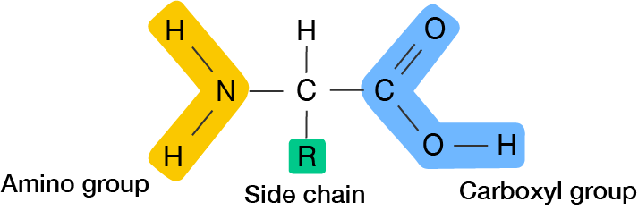
Proteins are polymer chains of amino acids, which are linked by peptide bonds. Amino acids have an amino (\(\ce{NH2}\)) group, a carboxyl (\(\ce{COOH}\)) group and a unique side chain (R group) which gives the amino acid its properties. There are \(20\) standard amino acids that combine in different sequences to form proteins. General structure of an amino acid
Proteins are essential for bodily functions like building and repairing tissues, facilitating chemical reactions and supporting the immune response. They are composed of carbon (\(\ce{C}\)), hydrogen (\(\ce{H}\)), oxygen (\(\ce{O}\)) and nitrogen (\(\ce{N}\)) atoms.
The 3D structure of a protein is very important for its function, so it is important to understand how we arrive at its final shape. Proteins are organised into four levels of structure:
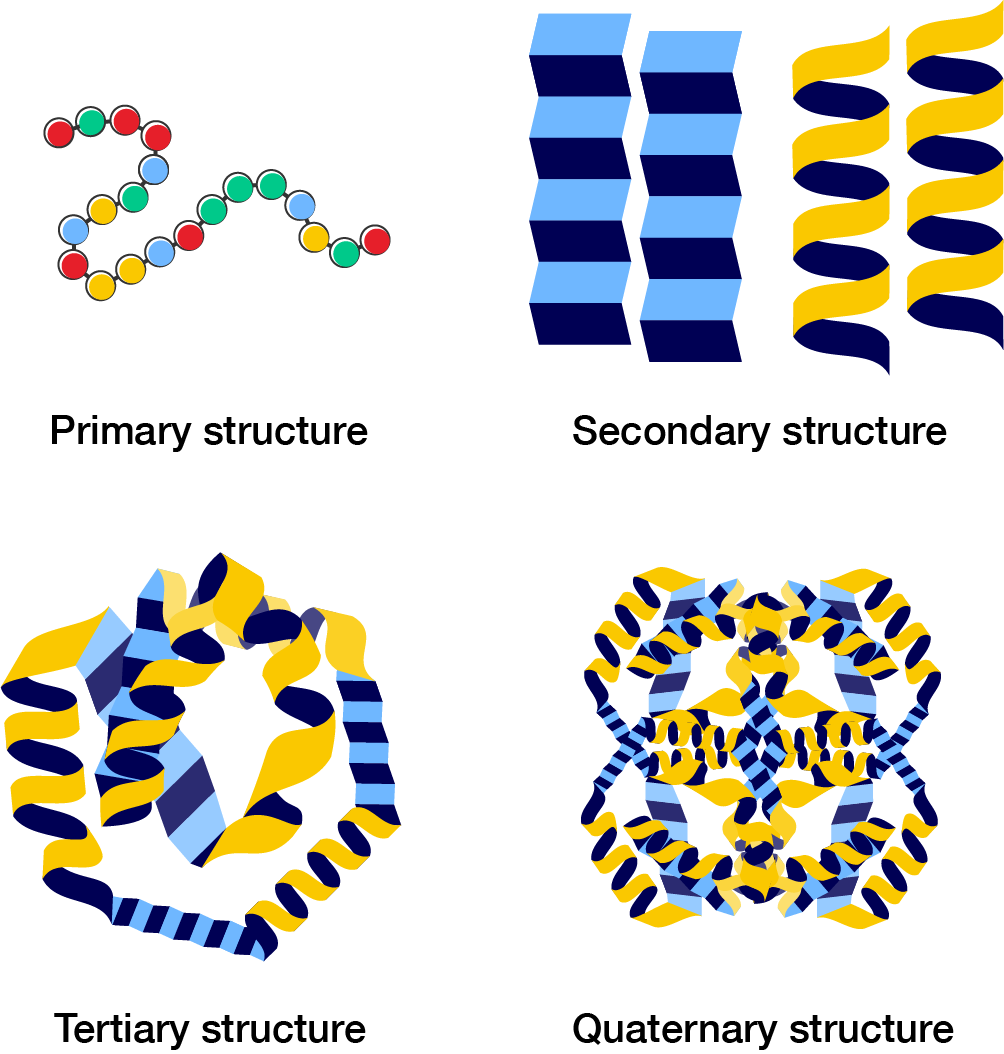
Primary structure: the linear sequence of amino acids linked by peptide bonds
Secondary structure: the alpha helices or beta pleated sheets formed by hydrogen bonds between amino acid side chains
Tertiary structure: the overall 3D shape of a single protein molecule created by disulfide bonds, hydrogen bonds, ionic bonds and hydrophobic interactions between amino acid side chains
Quaternary structure: the arrangement of multiple protein subunits into a more complex structure. Not all proteins have a quaternary structure.
Primary, secondary, tertiary and quaternary structures of proteins
Structure of proteins
Primary structure (top left): coloured balls connected in a chain.
Secondary structure (top right): two corrugated sheets which are beta pleated sheets or two helical shapes which called alpha helices.
Tertiary structure (bottom left): beta pleated sheets and alpha helices connected together in a tangled chain.
Quaternary structure (bottom right): four units of the tertiary structure.
An example of a protein is haemoglobin, which transports oxygen in the blood. It is able to bind oxygen-carrying heme molecules because of its complex quaternary structure. Use the 3D model shown to look at the structure of haemoglobin.
Four protein subunits (coloured green, light blue, pink and yellow) are represented using coils of ribbon. Each subunit contains a heme molecule with its full structure shown using ball-and-stick models. Each heme molecule interacts with iron, represented by an orange sphere. Each iron interacts with an oxygen molecule, shown using its ball-and-stick model. All interactions are shown using dashed white lines.
Formation of proteins
Eggs are a great source of protein and amino acids, by Conger Design via Pixabay, licensed under CC0
High protein foods include meat, eggs and beans. When we eat these foods, our bodies break down the proteins into amino acid building blocks, which we can then use to synthesise proteins required by our bodies.
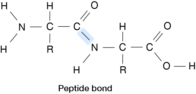
Like carbohydrates, the peptide bonds are between amino acid monomers through condensation reactions. Peptide bond
However, the overall process for protein synthesis is a bit more complex. Go to protein synthesis to learn more.
Lipids
Lipids are hydrophobic ("hydro" meaning "water" and "phobic" meaning "hating") macromolecules, including fats, oils and steroids. They are composed of primarily of carbon (\(\ce{C}\)), hydrogen (\(\ce{H}\)) and oxygen (\(\ce{O}\)) atoms, but can contain other atoms.
Lipids are essential for storing energy, forming cell membranes, insulation, producing hormones and serving as signalling molecules. Unlike proteins and carbohydrates, lipids don't have a standard monomer but are often made up of fatty acids and glycerol.
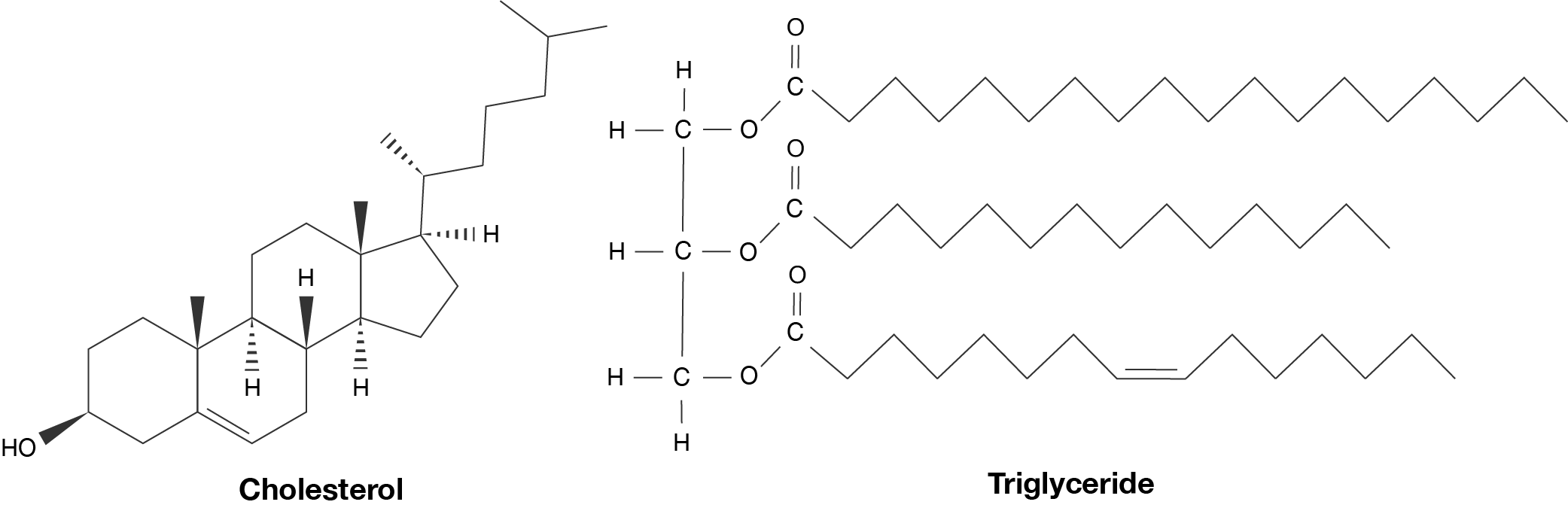
Common examples include triglycerides which store energy in fat cells, phospholipids which form the protective outer layer of cells, and cholesterols which help your body create bile. Cholesterol (left) and a triglyceride (right)
Formation of lipids
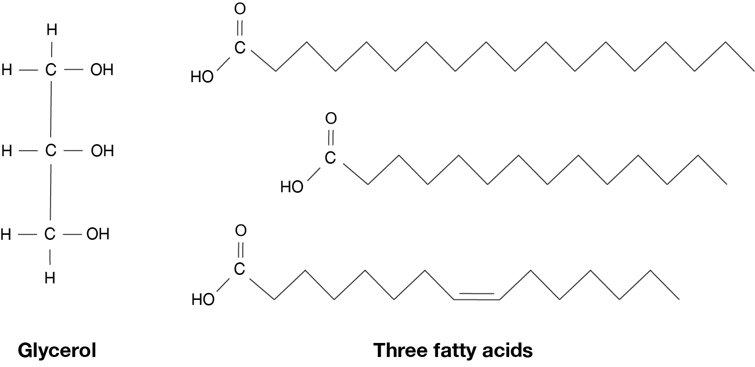
Lipids are diverse in their structure. Triglycerides and phospholipids form from condensation reactions between a mixture of glycerol, fatty acids and phosphate groups. Cholesterols, on the other hand, have a more rigid structure and form through more complex reactions. Glycerol (left) and fatty acids (right)
Just like proteins and carbohydrates, lipids can be obtained from foods we eat, like butter, neutral oils, fish, avocado, fatty meats and cheese. Cheeses are rich in triglycerides and other lipids, by Waldemar via Unsplash
Nucleic acids
Nucleic acids are macromolecules that store genetic information. The two main types of nucleic acids are:
deoxyribonucleic acid (DNA) which carries genetic instructions for the cell
ribonucleic acid (RNA) which plays a role in translating DNA into proteins.
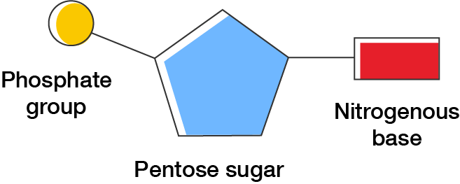
General structure of a nucleotide
Nucleic acids are composed of nucleotide monomers. These consist of a sugar, phosphate group and nitrogenous base.
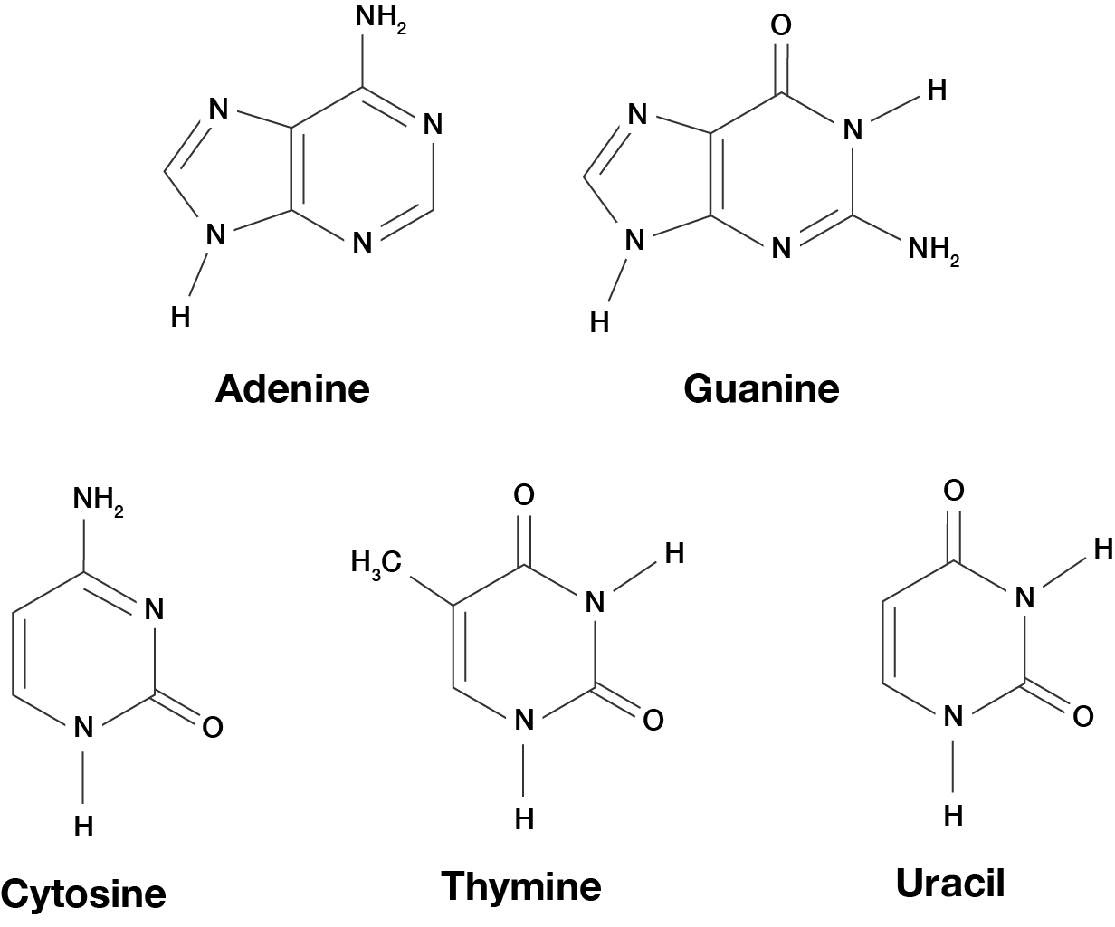
The bases are adenine, thymine (or uracil in RNA), cytosine and guanine. These four bases form pairs to encode genetic instructions for the cell. The atoms in these macromolecules are carbon (\(\ce{C}\)), hydrogen (\(\ce{H}\)), oxygen (\(\ce{O}\)), nitrogen (\(\ce{N}\)) and phosphate (\(\ce{P}\)). The nitrogenous bases
You will learn more about how DNA is transcribed into RNA, then translated into proteins in protein synthesis.
Summary
The features of the four types of macromolecules are summarised in the table.
Macromolecule
Monomer
Composition
Function
Examples
Carbohydrate
Monosaccharides
\(\ce{CHO}\)
Cell metabolism and function
Sugar, starch, fibre
Protein
Amino acids
\(\ce{CHON}\)
Tissue development and repair, chemical reactions, immunity
Hemoglobin, collagen, antibodies
Lipid
Not a polymer, but can contain glycerol, phosphate, fatty acids
Primarily \(\ce{CHO}\)
Energy storage, cell membrane component, communication between cells
Cholesterol, phospholipids, triglycerides
Nucleic acid
Nucleotides
\(\ce{CHONP}\)
Storage and expression of genetic information
DNA, RNA
Exercise
See how well you understand macromolecules and their characteristics with a quick quiz.