Building DNA
Use this simulation to practise building your own double-stranded DNA. This is a good way to make sure you understand how the base pairings work! You can also see DNA replication in action.
For an organism to develop, repair tissues and stay healthy, its cells must reproduce. You need a good understanding of cell reproduction in medicine for studying cancer and developing treatments, and in agriculture for improving crops. Use this resource to explore the replication of DNA and the process of meiosis.
Cell reproduction is how cells divide to create new cells, allowing for growth, repair, and continuation of life. It includes processes like:
Together, these processes ensure healthy development and reproduction. You have already learned about mitosis, so let’s discuss DNA replication and meiosis.
DNA replication is the process by which a cell makes an exact copy of its DNA before cell division. This occurs during interphase.
Just like mRNA transcription and protein translation, DNA replication follows the three main steps: initiation, elongation and termination.
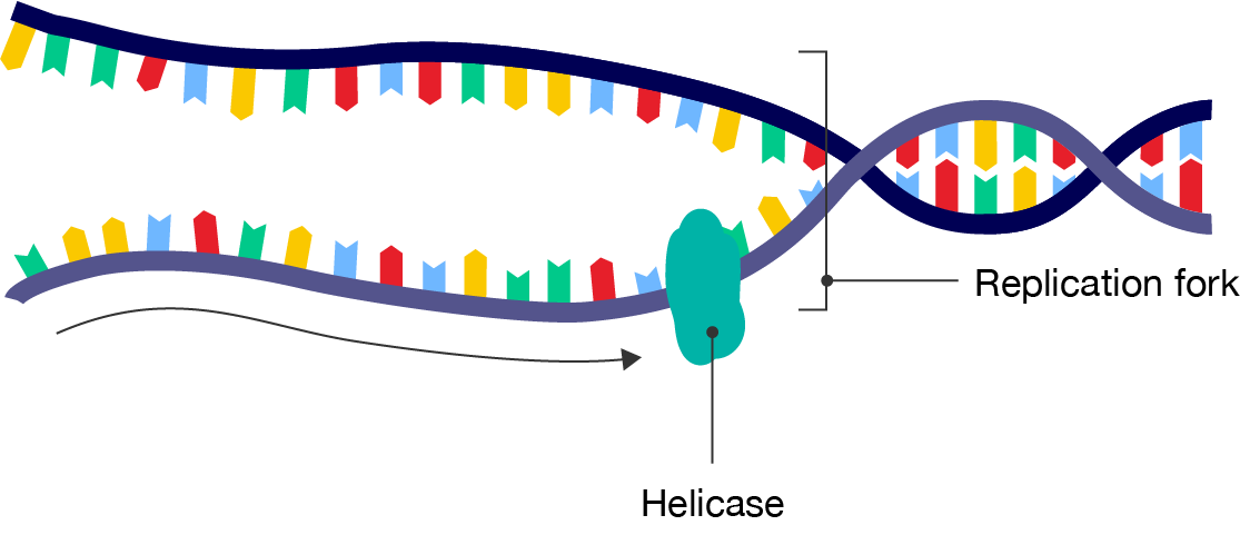
During initiation, an enzyme called helicase unwinds the DNA double helix and “unzips” the strands. These strands serve as templates for building new complementary strands.
Proteins attach to the DNA to keep the strands apart. This forms a replication fork, where two single strands of DNA are exposed. The initiation step prepares DNA for replication.
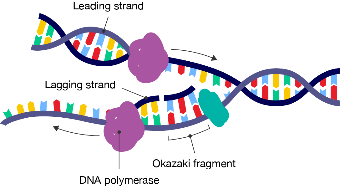
During elongation, DNA polymerase creates a new DNA strand by adding nucleotides that are complementary to the template strand. Unlike mRNA transcription, the base pairings are:
The strand that points towards the replication fork is called the leading strand. Nucleotides are added to the leading strand in the 5’ to 3’ direction, just like how the mRNA template is read from 5’ to 3’.
The strand that points away from the replication fork is also read in the 5’ to 3’ direction. But because the double helix continues to unzip as nucleotides are added, the DNA is replicated in short segments called Okazaki fragments. This strand is synthesised backwards and is therefore given the name lagging strand. The Okazaki fragments are joined together by the enzyme DNA ligase, forming a continuous DNA strand.
Once the new strands are formed, DNA polymerase checks for errors. The precise duplication of genetic material is crucial for the maintenance and function of living organisms.
If no mistakes are found, each template strand and new DNA strand are wound into a double helix with the help of DNA ligase. The result is two DNA molecules that consist of one new strand and one old strand.
Watch this video to see DNA replication in action.
Here, the DNA to be copied enters the complex from the left. One new strand is leaving at the top of frame and the other new strand is leaving at bottom.
The first step in DNA replication is the separation of the two strands by an enzyme called helicase. This spins the incoming DNA to unravel it: at ten thousand RPM in the case of bacterial systems. The separated strands are called three prime and five prime, distinguished by the direction in which their component nucleotides join up.
The 3 prime DNA strand, also known as the leading strand, is diverted to a DNA polymerase and is used as a continuous template for the synthesis of the first daughter DNA helix.
The other half of the DNA double helix, known as the lagging strand, has the opposite 3 prime to 5 prime orientation and consequently requires a more complicated copying mechanism. As it emerges from the helicase, the lagging strand is organised into sections called Okazaki fragments. These are then presented to a second DNA polymerase enzyme in the preferred 5 prime to 3 prime orientation. These sections are then effectively synthesised backwards.
When the copying is complete, the finished section is released and the next loop is drawn back for replication. Intricate as this mechanism appears, numerous components have been deliberately left out to avoid complete confusion.
The exposed strands of single DNA are covered by protective binding proteins. And in some systems, multiple Okazaki fragments may be present. The molecular reality is very different from the iconic image of the double helix neatly separating into two DNA copies as so often depicted.
Meiosis is a special type of cell division that produces sex cells called gametes, such as sperm and eggs. Gametes are haploid cells, meaning that they have a single set of chromosomes. Compare this to mitosis, which produces diploid cells with two sets of chromosomes.
Meiosis is crucial for sexual reproduction—that is, reproduction involving two parents—ensuring that offspring receive a consistent number of chromosomes.
This process involves two divisions and results in four genetically unique daughter cells.
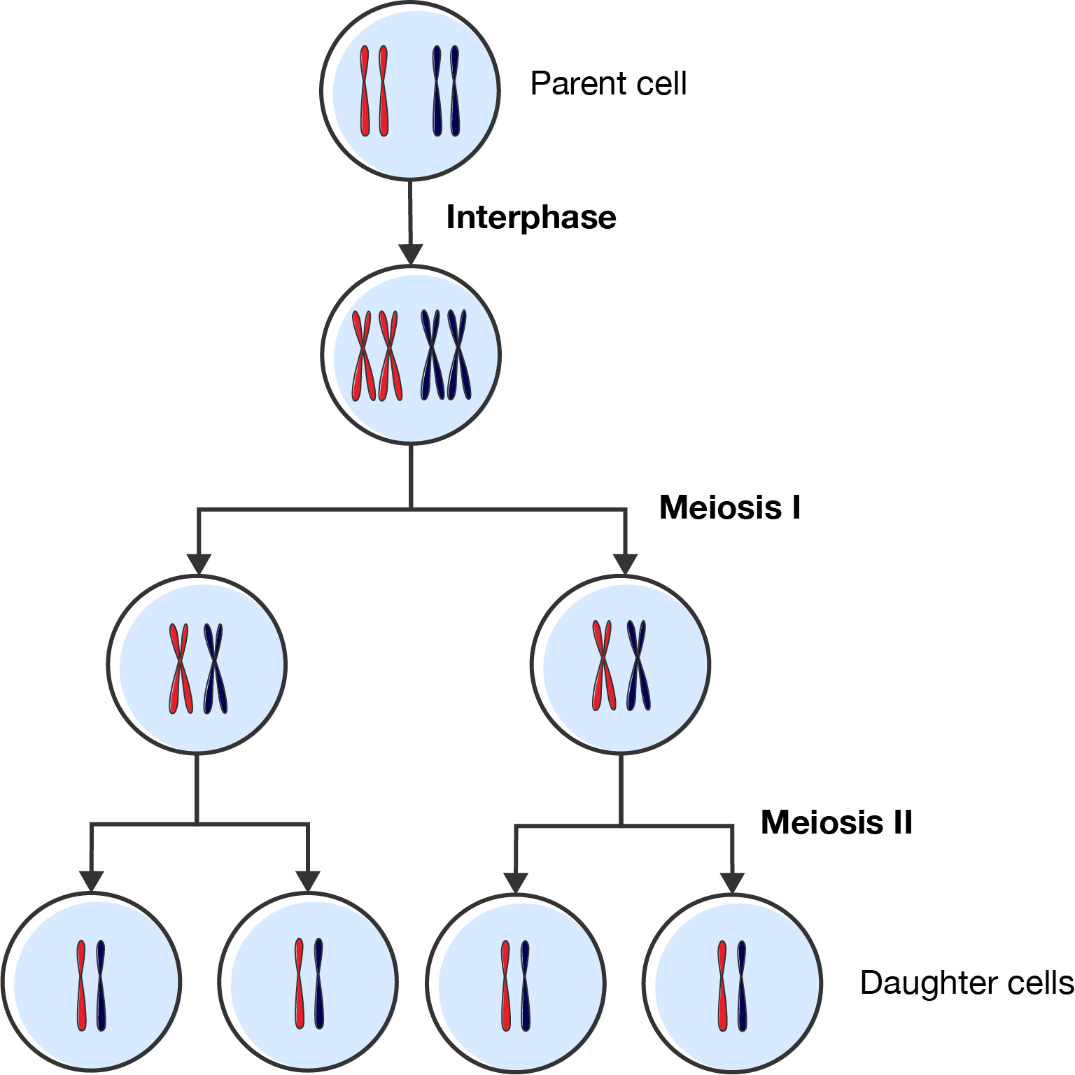
The first division is called meiosis I. This begins after interphase, when the DNA has been replicated. The stages of meiosis I are:
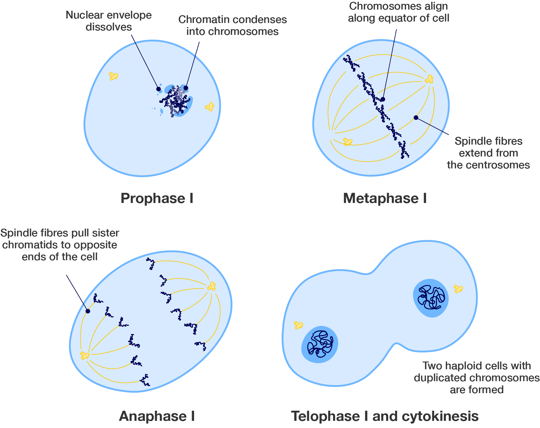
At the end of meiosis I, there are two haploid cells with duplicated chromosomes.
The second division is called meiosis II. The stages of meiosis II are:
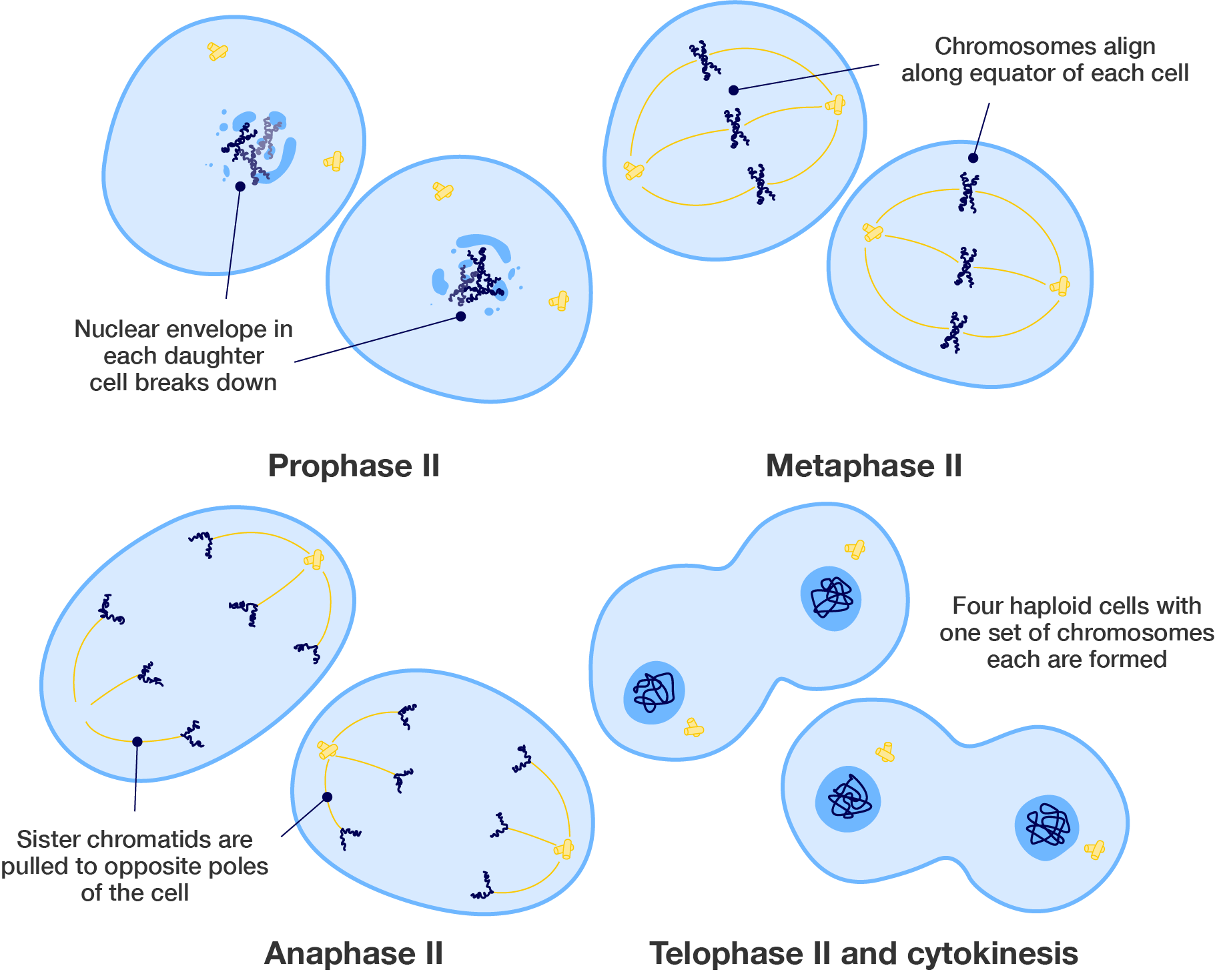
At the end of meiosis II, there are four haploid cells, each with single chromosomes.
Meiosis makes sure there is genetic diversity. Watch this video to see meiosis in action.
First, it's good to know that there are two types of cell division processes.
The simpler one is mitosis which produces two identical cells with exactly the same genetic information. You can think of them as clones of each other. And to find out more, why not look at our videos about mitosis?
The other process is meiosis, is a much more complicated process creating not 2 but 4 cells with only half the number of chromosomes, and crucially, all genetically different from each other.
Both mitosis and meiosis include the same phases: prophase, metaphase, anaphase and telophase. Except, in meiosis, they happen twice, so they usually referred to as one and two.
The easiest way to remember these phases' names is to remember IPMAT. So let's look at meiosis in more detail.
As always, cellular division starts with a process called DNA replication. This involves making two identical copies of the original DNA molecule. The cell ends up temporarily with double the normal number of chromosomes. You can learn more about DNA replication in this video.
In prophase 1, the duplicated chromosomes join up with the pair from the other parnent. So the mother's pair bind with the parther's pair, forming a group of two chromosomes called homologous chromosomes.
What happens next is vital for successful meiosis. As each chromosome is lined up next to its partner's pair, one chromatid from each side gets entangled with a corresponding chromatid from the other side. This is called crossing over.
During this brief period, the two chromatids swap certain sections of DNA. This is called recombination. The sections that they trade correspond to the same location so that each chromatid retains the correct number of genes.
Recombination is really important because it creates variety. The new cells aren't identical to the other parents and they also are different to one another as well. They are new genetic combinations.
In fact, that's the whole point of sexual reproduction. It increases genetic variability.
Each chromatid is now different and as each one will end up in a separate gamete, it means each sex cell is genetically different from all others. This explains why brothers and sisters are different despite having the same appearance. Only identical twins have the same genetic makeup as they both originate from the exact same egg and sperm.
Now, back to meiosis. Next comes metaphase 1 as the chromosomes align themselves up in the middle of the cell. And in anaphase 1, the spindle fibres pull the chromosomes apart to opposite ends. And during telophase 1 and cytokinesis, the cell pinches apart in the middle and the nuclear membrane reforms around the two new daughter cells. That's the end of meiosis 1.
Now, for part 2. We start with our recombined daughter cells, each still with 46 chromosomes,. But sperm and egg cells only have 23 chromosomes, so we need to cut these cells in half.
Here goes a second round of division. The process is exactly the same as before, except that there is no DNA replication. We start straight with prophase 2 with chromatin clumping again to form chromosomes, then align in the middle of the cell during metaphase 2 and chromatids are pulled apart during anaphase 2 by the spindle fibres. Telophase and cytokinesis pinch the cells together with four new granddaughter cells being formed.
The end of meiosis gives us four different sex cells each, each with only 23 chromosomes ready for future fertilisation. But that's a story for another video.
If you like this video, give it a thumbs up and don't forget to subscribe. Comment below if you have any questions. Why not check out our Fuse School apps as well. Until next time!
See how well you understand cell reproduction with a quick quiz.
Images on this page by RMIT, licensed under CC BY-NC 4.0
Building DNA
Use this simulation to practise building your own double-stranded DNA. This is a good way to make sure you understand how the base pairings work! You can also see DNA replication in action.
Cell division: Mitosis and meiosis
Use this interactive tutorial to learn about mitosis and meiosis, and compare them!
Mitosis and meiosis simulation
Explore the difference between mitosis and meiosis by watching them happen side-by-side.


RMIT University acknowledges the people of the Woi wurrung and Boon wurrung language groups of the eastern Kulin Nation on whose unceded lands we conduct the business of the University. RMIT University respectfully acknowledges their Ancestors and Elders, past and present. RMIT also acknowledges the Traditional Custodians and their Ancestors of the lands and waters across Australia where we conduct our business - Artwork 'Sentient' by Hollie Johnson, Gunaikurnai and Monero Ngarigo.
More information